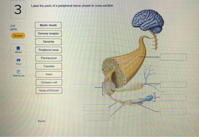
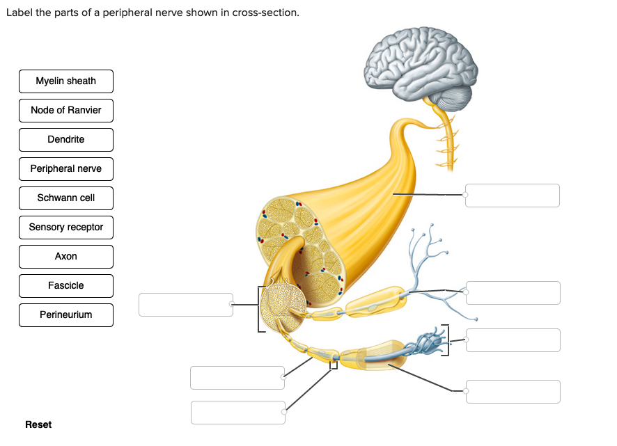
Label the Parts of a Peripheral Nerve Shown in Cross-section.
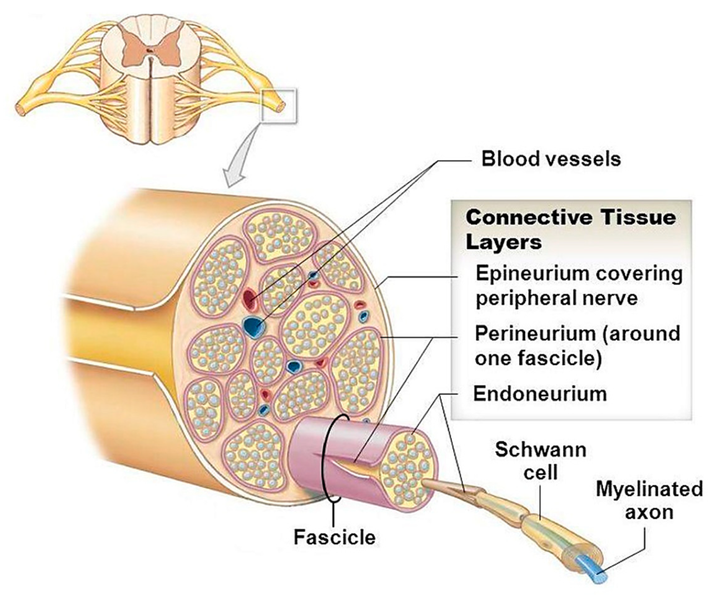
Peripheral nerves consist of bundles of myelinated and non-myelinated nerve fibers enveloped by connective tissue. A nerve provides a structured pathway that supports the electrochemical nerve impulses transmitted along each of the axons.
Applied Sciences Free Full Text Bioactive Glasses And Glass Polymer Composites For Neuroregeneration Should We Be Hopeful Html
Cross section through the thalamus.
. Learn vocabulary terms and more with flashcards games and other study tools. Nerve a bundle or bundles of nerve fibers. Can be inflamed in autoimmune rheumatologic disorders.
For each cranial nerve place the label in the appropriate box categorizing it based on function. Up to 24 cash back Their fibers cross over in the spinal cord. The dense pink-colored band is the perineurium.
The latter of which can either be sympathetic fight or freight response or. Learn cross section nerve with. Label the parts of a peripheral nerve shown in cross-section.
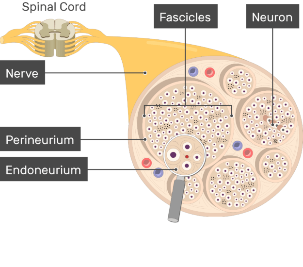
The peripheral nervous system PNS consists of all the nerves branching out of the brain and spinal cord the central nervous system CNS. In the transverse section find epineurium perineurium and. A layer of connective tissue called the perineurium pn surrounds each fascicle.
The spinal cord which consists of the major nerve tract of vertebrates runs down from the bottom of the brain through the passageway of the spinal column. Wavy tissue poorly stained because of the high lipid content of the myelin axons surrounded by myelin sheaths. The 40X image shows a cross section through four fascicles f that are part of a nerve.
On glass slide 51 in your Histology slide box longitudinal and transverse sections of a sciatic nerve from a horse are evident triple stain. A nerve is an enclosed cable-like bundle of axons the projections of neurons in the peripheral nervous system PNS. Nerves are composed of more than just nervous tissue.
This is a Peripheral Nerve cross-section please use ARROWS to label the following. Schwann cell myelin sheath Nucleus of Schwann cell. Therefore ganglia can be distinguished from peripheral nerves by the presence of neuronal cell bodies.
In the central nervous system the analogous structures are known as tracts. This is a peripheral nerve seen in cross-section. This area is made up of all the nerve fibers that direct the reflex actions and convey the impulses that go back and forth to the brain.
Midbrain _ crevical nerves _ thoracic nerves _ lumbar nerve. Bundles of axons in the PNS are referred to as nerves. The central nervous system is comprised of the brain and spinal cord while the peripheral nervous system includes all spinal and cranial nerve fibres providing end organ innervation.
These structures in the periphery are different than the central counterpart called a tract. The last pair of ascending pathways are the ventral and dorsal spinocerebellar tracts. Axons are neuronal processes specialized for electrical impulse conduction.
Label the parts of a peripheral nerve shown in cross-section. Label the parts of a peripheral nerve shown in cross-section. Learn cross section nerve with free interactive flashcards.
At the periphery of the fascicle observe the perineurium made up of several layers of flattened cells. The star of the show brain is easily recognizable because it appears highly convoluted full of ridges gyri and indentations sulciThe paired thalami appear as two circular masses in the midline forming the walls of the third ventricleThe neurocranium appears as a. Choose from 5000 different sets of cross section nerve flashcards on Quizlet.
The dense pink-colored band is the perineurium. Myelinated Nerve with Endoneurium and Perineurium cross section. If you imagine the CNS as the main highway then the PNS forms all the connecting secondary roads.
Most of the fibers in these pathways transmit pain temperature and coarse touch impulses sensations that we are aware of but are difficult to localize precisely on the body surface. Start your trial now. Note the presence of neuronal fascicles each of which is a bundle of nerve fibers and note that the organ itself is a bundle of fascicles.
Diagram of a peripheral. Start studying Peripheral nerve slide- Cross-Section. Of note the peripheral nerve is also further subdivided into the somatic and autonomic divisions.
Spinal Cord Cross Section. They have connective tissues invested in their structure as well as blood vessels supplying the tissues with nourishment. It is a highly specialized layer that acts as a barrier and protects the nerve from the environment.
Ganglion clusters of neuronal cell bodies in the peripheral nervous systems as well as associated glial cells and axons. Start studying Histology cross section of a peripheral nerve. Between the axons you will see delicate connective tissue and an occasional fibroblast which constitute the endoneurium.
These allow electrical impulses to travel to and from the furthest regions or periphery of the human body. The spinal cord which consists of the major nerve tract of vertebrates runs down from the bottom of the brain through the passageway of the spinal column. Select View 50 um T.
Diagram Orienting yourself within such a cross section is easy. This is a peripheral nerve seen in cross-section. This is peripheral nerve.
The corpora quadrigemina cerebral peduncles and red nucleus are parts of the __ of the brainstem. A whole nerve is surrounded by connective tissue called the epineurium not shown by this slide. In the peripheral nervous system axons are bundled together in structures called nerves.
Learn vocabulary terms and more with flashcards games and other study tools. In the transverse section find epineurium perineurium and endoneurium see Figs.
Solved Label The Parts Of A Peripheral Nerve Shown In Chegg Com

The Peripheral Nervous System Anatomy And Physiology
Solved Label The Parts Of A Peripheral Nerve Shown In Chegg Com
Nerve Structure Anatomy


Comments
Post a Comment